- Orthogenetic Surgery
-
Orthogenethic Surgery
Orthogenethic Surgery is to correct the conditions of the jaw and face, to correct disorders of the jaw, face, sleep problems and malocclusion. It can also be referred to as corrective jaw bone surgery.
Orthogenethic surgery is always a planned or elective surgery and it is mandatory for it to be performed by an expert surgeon, qualified and experienced in all the techniques of the surgery.
Modi's dental Hospital / clinic has a team of Oral and Maxillofacial surgeons who are highly experienced in performing complicated surgeries like Orthogenetic Surgery. The team has performed many surgeries till date and all of them have been successful.
Modi's Dental clinic / Hospital’s expertise in this field has been supported by use of modern technology, techniques and a dedicated team of super specialist Doctors.
Modi's Dental Clinic, over the years has earned a reputation for being a famous centre for Jaw Correction surgeries with its Super Specialist Team of Doctors and successful outcomes. They are capable of handling all kinds of treatment procedures in any of the super specialty in dentistry, thanks to their infrastructure and expertise in the respective fields.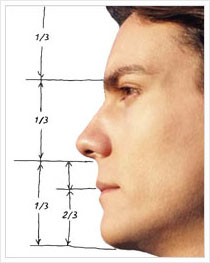
- Reduction Jaw Surgery
-
Jaw Reduction Surgery
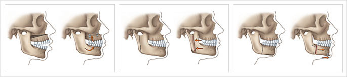
Jaw reduction is a type of aesthetic plastic surgery in which the objective of treatment is to narrow the lower one-third of the face - particularly the contribution from the mandible and its muscular attachments. There are several techniques for treatment - including surgical and non surgical methods.
Facial aesthetics
A square lower jaw is generally considered a very masculine trait. Widening of the lower third of the face can cause either a square appearance to the face as a whole or can distort the natural appearance of the angle between the chin and the neck.
Whereas square lower jaws are often considered a positive trait in Caucasian and many Asian men, a wide mandible can cause significant facial discordance and/or masculinization of the female face-particularly in those of East Asian descent. Even in certain men, the size of the lower jaw can cause facial disharmony-particularly when there is asymmetry.
A wide lower face can primarily be caused by enlargement of the mandible or masseter muscle.
Causes
The primary cause of an enlarged mandible is developmental or congenital. There are some rare disorders that can further widen the jaw such as acromegaly.
Conversely, while a masseter muscle can be large due to congenital reasons, it can commonly be an acquired deformity. Like any muscle it increases in size with exercise. Behaviors such as repeated gum chewing, teeth clenching, or bruxism can contribute to enlargement of the muscle.
Techniques
There are several jaw reduction techniques available - both surgical and non-surgical. Ideally prior to selection of a treatment, the patient is examined to determine whether the cause is due to the bone, the masseter or both. Additionally, if a treatable cause is present it should be identified.
Surgical reduction
Surgical techniques are used to directly reduce the size of an enlarged mandible. Incision can be to the inside or outside of the mouth, though the internal incision is the most common because it leaves no visible scar. A bur is used to remove the outer layer of the enlarged mandible, narrowing the jaw
Potential complications include injury to the inferior alveolar nerve which provides sensation to the lower lips and teeth. - Distraction Jaw Surgery
-
Distraction Osteogenesis in Maxillofacial
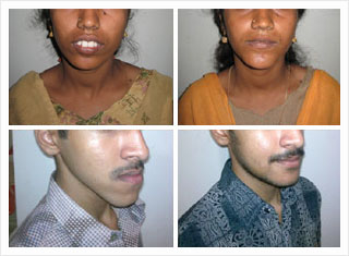
Maxillofacial Surgeons use this technique for the correction of micrognathia, midface, and fronto-orbital hypoplasia in patients with craniofacial deformities.
Correcting the majority of congenital craniofacial defects, as well as some facial injuries resulting from trauma, requires making bones longer. Distraction osteogenesis is an effective way to grow new bone, but it is much more difficult to accomplish in the face than in other areas of the body. Bones must often be moved in three dimensions, as opposed to just one, as in a limb, and scarring must be kept to a minimum. Researchers are attempting to improve the distraction devices used in the face. Until recently, the mechanisms were external and only operated along straight lines. Now, maxillofacial surgeons can use curvilinear devices capable of moving bone in three dimensions.
These new devices still need to be improved. They depend on patient caretakers reliably turning a screw. The next goal is to create devices that will move bone continuously, in daily increments of 1 mm. These continuously moving devices would cause less pain, wouldn't require daily patient compliance, and might promote faster bone growth. At the moment, researchers are testing a continuously moving device in animal models, and they have found that the device's components are durable, that its user interface works, and that it is tolerated by the body. When the position sensor in the device is perfected, the device will be ready to use in people.
In distraction osteogenesis procedures involving the face, it is critical that bone movements be carefully planned before a device is implanted. No existing device is capable of changing its trajectory mid-course, and small skeletal changes lead to large changes in the structure of the face. Recently researchers have developed state-of-the-art software capable of simulating the entire process of distraction osteogenesis. The 3-D planning tool uses data from CT scans to create a segmented model of the patient's skull, and it then calculates the vector of movement required to achieve desirable bone positioning. Outcome of CT scans can be overlaid on the original model to assess the effectiveness of the procedure. In the future, researchers hope that the distraction devices used in maxillofacial procedures will continue to improve, along with the corresponding software.
RESULTS: All patients achieved lengthening of their jaws. However, premature consolidation was noted in two patients, and one patient had significant relapse. - Genioplasty
-
Genioplasty Chin Surgery
Genioplasty (Mentoplasty) Malar Augmentation Or Reduction (Chin Surgery)
The chin provides harmony and character to the face. A strong chin or prominent jaw line is considered to be aesthetically pleasing, especially in males. When chin surgery is indicated, whether by anterior horizontal mandibular osteotomy (AHMO) or by alloplastic implant augmentation, it can create an aesthetically pleasing facial contour and establish proportionate facial height. In addition, the AHMO can improve obstructive sleep apnea by elevating the hyoid bone.
Most genioplasty procedures are done to improve the mandibular profile in order to obtain a more natural profile. Genioplasty can shorten or lengthen the lower third of the face. Facial asymmetry may be corrected by rotation of the chin-point to coincide with the midline. The advantages of osseous genioplasty are versatility, reliability and consistency in correcting problems in the sagittal and vertical planes to achieve greater chin projection. In order to be able to make an appropriate recommendation, the correct preoperative workup should be performed, including soft and hard tissue analyses. Ideally, cephalometrics and video cephalometric predictions would also be performed. Anatomy and Analyses
It is important for the surgeon to be familiar with the classic soft tissue analysis and diagram of facial proportions. The size, shape and position of soft and hard tissue can enhance facial harmony and symmetry. The relationship between soft tissue and bone is important for planning the bony chin correction. For chin advancement, the bone to soft tissue proportion is 1:0.8, meaning that 1 mm of bony change is associated with 0.8 mm of soft tissue change.
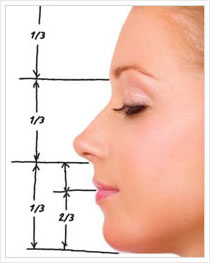
The face can be divided into upper, middle and lower thirds. The upper third of the face spans from the hairline to the glabella; the middle third from glabella to subnasale; and the lower third from subnasale to menton. The lower third of the face can be further divided into an upper half (Sn to vermilion of the lower lip) and a lower half (Me to vermilion of the lower lip). The face is "balanced" when the three thirds are of similar height. Cephalometric analysis ensures that skeletal and occlusal disparities are identified and can be corrected before or at the same time as a genioplasty. Many patients that complain of a small chin truly do not have microgenia.
They often have a true deficit of the mandible in the sagittal plane, which can be a class 2 malocclusion (retrognathia) or normo-occlusion (retrogenia). Retrognathia is ideally corrected with a bilateral sagittal split osteotomy (BSSO); however if the discrepancy is small, advancement genioplasty may sufficiently camouflage the facial profile into an orthognathic appearance. Retrogenia (chin point deficiency in the setting of a class I occlusion) and mild retrognathia (<3 mm) are ideal cases for a genioplasty. It is important to understand the relationship of the dentition to the chin point. The boney chin point should be about 2 mm posterior to the labial surface of the mandibular incisors. This will help maintain a natural labiomental fold.
The position of the labiomental angle is paramount and profoundly influences the aesthetic outcome. Cephalometric analysis helps the surgeon to plan the operative procedure. The treatment plan is based on incorporation of these data into clinical assessment that will facilitate a postsurgical profile that is esthetically pleasing. Perceived Chin Abnormalities Due to Anomalies of The Maxilla
When facial analysis identifies disharmony within a patient's profile, the surgeon must determine whether there is an underlying occlusal and skeletal deformity or merely a poorly or over-projected mentum. True maxillomandibular discrepancies should be addressed with orthognathic surgery.
In the case where occlusion is stable and a small mandibular deficiency exists (retrogenia), an isolated mandibular sagittal deficiency may be a candidate for an AHMO.
To highlight the importance of the correct diagnosis, one can take the common occurrence of a patient complaining of a "small chin." A recessed chin may be retrogenia or microgenia. An over projected chin may be macrogenia or prognathia. Micrognathia and macrognathia are rare. Prognathia and retrognathia more commonly contribute to chin point abnormalities.
In the setting of a patient complaining of a small chin, the lateral profile should be evaluated. Concavity or convexity in conjunction with the proportions of the middle and lower third of the face should be considered in the planning. The maxilla should be evaluated. If the maxilla is set appropriately in the sagittal plane and there is mild retrognathia (<3 mm) or retrogenia, then a genioplasty is appropriate. However, if a maxillary developmental dysplasia is present, a formal orthognathic work-up should be done.
In contrast, patients complaining of a "prominent chin" often have pseudomacrogenia. These individuals may have maxillary sagittal hypoplasia, which manifests with a retruded upper lip, a midfacial concavity or deficiency, and a chin that may appear prominent in the sagittal plane. Since the true etiology is maxillary hypoplasia, the corrective procedure would be a Le Fort I osteotomy to advance the maxilla anteriorly to coincide with the chin point. A pitfall would be for a novice surgeon to perform a genioplasty to set the chin point back to coincide with the maxilla.
In maxillary vertical deficiency, the patient presents with pseudomacrogenia due to the counterclockwise rotation of the mandible. In this case, the chin is accentuated and appears larger than normal. Patients with this condition have a short lower third facial height and present with poor maxillary tooth show at rest and when smiling. When the mandible is placed in the normal centric relation, the chin point increases in the sagittal plane. Maxillary vertical height correction will allow for a more natural position of the chin and only then can a decision be made on the need for genioplasty.
Maxillary vertical excess may manifest as pseudomicrogenia due to the excessive downward growth of the maxilla causing a clockwise rotation of the mandible. In such cases, the rotation of the mandible results in the appearance of a small chin due to poor projection of the chin in the sagittal plane. The patient will likely have excess gingival show, a long lower third facial height and mentalis muscle strain from the forces needed to close the interlabial gap. The treatment for this type of facial anomaly may be to reposition the maxilla superiorly, particularly in the posterior area.
Maxillary sagittal hyperplasia is extremely rare. Patients may complain of a small chin as well. Once again, this is most likely a case of pseudomicrogenia, where the chin appears relatively small due to the prominence of the maxilla in the sagittal plane. These patients will have a convex facial profile associated with maxillary protrusion and an acute nasolabial angle. This form of microgenia can be corrected with repositioning of the maxilla, after which a decision can be made on the need for an adjunct genioplasty. Evaluation of The Mandible
After a thorough investigation to rule out any maxillary discrepancies, the next step is to evaluate the mandible. For a patient with mandibular hypoplasia with either gross malocclusion or severe hypoplasia (greater than 4 mm), a formal orthognathic work-up is necessary. The risks of advancing or augmenting a chin greater than 5-6 mm include an unnatural appearance, a deep labiomental angle, and the risk of advancing the chin point past the lower central incisor. Severe mandibular sagittal hypoplasia is corrected with a BSSO, and genioplasty should be viewed as an adjunct procedure. Prognathia in the setting of class 3 malocclusion should be corrected by a setback with a BSSO. Isolated true macrogenia in the presence of a normal class 1 occlusion can easily be treated with a genioplasty, setting the chin point back and even reducing chin height if needed. Indications for Isolated Genioplasty (Mentoplasty)
After careful scrutiny of the skeletal, dental and soft tissue structures, there exist certain cases that are amenable to isolated genioplasty. An isolated genioplasty can be considered if functional occlusion is present and the lower third profile has mild hypoplasia or hyperplasia in the sagittal or vertical plane. A sagittal hypoplasia (3-4 mm) in the setting of functional normo-occlusion with acceptable facial proportions is an ideal candidate for AHMO with advancement. A variation of the standard sliding genioplasty is the "jumping" genioplasty. The "jumping" genioplasty is ideal for sagittal advancement when vertical reduction is needed.
Patients who are considered for isolated genioplasty should have a good overall profile and occlusion. The surgical goals for these patients include creating an aesthetically pleasing facial contour and establishing proportionate facial height. Ideal candidates for a genioplasty are: (1) retrogenia, i.e., recessed chin point with class I occlusion; (2) mild retrognathia (<4 mm) with a functional occlusion; and macrogenia.
For example, in a patient that may have a long lower third of the face, a reduction genioplasty is performed to reduce the vertical dimension of the chin. Vertical reduction is done by performing a second horizontal osteotomy that is parallel to the first osteotomy, and a segment of bone is removed. Another indication for an isolated genioplasty may be a mild asymmetry, when the chin does not coincide with facial midline. An oblique triangular wedge of bone can be removed from one side and transplanted to the other side to correct chin asymmetry.
In all cases, the chin has to be rigidly fixed by miniplates, wire or screws. A variety of genioplasty is the "jumping" genioplasty. This type of procedure allows the surgeon to both increase the chin projection and shorten the vertical dimension of the chin simultaneously. After the osteotomy is completed, the basilar segment is elevated on top of the upper symphysis. Genioplasty - Surgical Technique - Procedure
Genioplasty can be performed under local anesthesia with IV sedation or under general anesthesia. General anesthesia is more commonly used with this procedure.
Lidocaine with epinephrine is infiltrated along the depth of the buccal vestibule. An incision is made in the buccal vestibule, initially perpendicular to the mucosa then perpendicularly to the muscle and bone. The dissection is continued in the subperiosteal plane to identify the mental foramen on both sides. After identification of the mental foramen, the mental nerves should be protected from both direct and traction injury.
The osteotomy is done under the apices of the teeth and the mental nerve. Upon completion of the osteotomy, the chin is then rigidly fixated. A step-off (sharp edge) at the posterior part of the genioplasty should be avoided. The contour of the mandible should be smooth. There are a variety of techniques used for fixation of the chin including wires, resorbable or titanium bone plates. Closure should be done in multiple layers. The mentalis muscle must be reapproximated.
The muscle layer can also be reattached to the chin using Mitek anchors. We prefer using two Mitek anchors to secure the mentalis muscle to bone. Alternatively, simple reapproximation with two horizontal mattress sutures is acceptable. This prevents ptosis of the mentalis muscle. Nonfixation may result in a "witches chin." A compressive chin dressing is worn for 5 days postoperatively. The oral mucosa is closed with a running 3-0 chromic suture.
The advantage of this procedure is its versatility, reliability and reproducible correction of chin point discrepencies. The disadvantages, when compared to alloplastic augmentation, include increased operative time, bleeding and incidence of mental nerve hypoesthesia.
- Frenectomy
-
Frenectomy
Frenectomy is a surgical procedure that involves the removal of the ‘frenulum’, done mostly for orthodontic purposes, usually inside the middle of the upper lip or under the tongue. It is performed in both adults and children routinely by Dental Surgeons.
This procedure is performed by qualified and experienced Dental surgeons and requires a great degree of precision. At Modi's Dental Frenectomy is done routinely by highly qualified and experienced surgeons with very desirable outcomes.
Keeping pace with recent advances in Technology, Modi's Dental has trained Surgeons, who have adopted the latest ‘Laser Frenectomy’ technique also as a part of their array of procedures.
Modi's Dental Hospital / Clinic in its quest for perfection has emerged as the best centers for Frenectomy where one gets to see modern technology and infrastructure being used with a human touch. This blend of technology and expertise makes Modi's Dental Clinic / Hospital a premier one stop shop for all dental procedures routine and rare.
- Dental Alveoloplasty
-
Dental Alveoloplasty
Alveoloplasty is surgical procedure where the alveolar ridges are shaped and smoothened in preparation of denture fixation after removal of several teeth.
This is a super-specialty procedure and is often delicate. The procedure has to have a great degree of perfection and that is precisely what the surgeons at Modi's Dental are trained to do.
Modi's Dental Hospital / Clinics routinely get cases for Denture Fixation and in the preparatory phase Super Specialists at Modi's dental perform Alveoloplasty wherever required to ensure smooth fixation of Dentures in patients.
Modi's Dental Hospital / Clinics adoption of newer techniques and Technology in dentistry has helped them to successfully perform such procedures with relative ease. All this is backed by expertise of the Team of highly trained Doctors which have ensured that Modi's dental is successful and the best center for such procedures. - Impaction & Complicated Extractions
-
Impaction & Complicated Extractions
Impaction is defined as the failure of a tooth to erupt into the dental arch within the specified time. In case of discomfort or pain, the tooth will need to be extracted or exposed surgically.
Dental impaction is seen very often and Impaction surgeries are done routinely by Surgeons at Modi's Dental Hospitals / Clinics. Modi's Dental does all types of impaction surgeries like Disto-angular Impaction, Vertical Impaction and Canine Impaction.
Surgeons at Modi's Dental Hospitals / Clinics are well trained and experienced in handling all types of Impaction Surgeries and keeping up with Modi's dental’s motto they try and keep it as pain free as possible.
Equally important to the surgery is post surgical care and rehabilitation. Doctors at Modi's dental are trained to complete whatever surgery they have started which is Pre Operative sedation, Surgery or extraction, Suture, and post surgical care.
Modi's Dental is capable of handling all procedures and surgeries with the Infrastructure, equipment and trained Doctors and staff which make them the best dental care Centre.

Dental Surgeries
Copyrights ©2014: Modi's Dental Clinic - Responsive Medical and Health Template